


This material is intended for people without medical education who want to know more about osteochondrosis than what is written in popular publications and on the websites of private clinics.The patient asks the doctor a variety of specialist questions that characterize a complete misunderstanding of the topic of osteochondrosis.Examples of such questions include: "why does my osteochondrosis hurt?", "congenital osteochondrosis is found, what should I do?"Perhaps the apotheosis of such illiteracy can be considered a rather common question: "Doctor, I have early signs of chondrosis, how scary is it?"This article aims to compile material on osteochondrosis, causes, manifestations, methods of diagnosis, treatment and prevention, and answer the most frequently asked questions.Since all of us, without exception, are patients with osteochondrosis, this article will be useful to everyone.
What is the structure of the intervertebral disc?
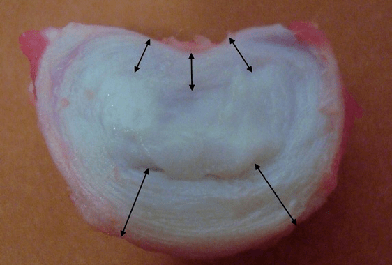
Each intervertebral disc consists of two distinct parts:
- outer fibrous ring, composed of dense fibers that cover the disc from the outside around the perimeter;
- the internal elastic component is the nucleus pulposus.
The fibers of the annulus fibrosus are very dense and elastic.Over the years, elasticity is lost, and at the age of 60 the fibrous ring becomes rigid.Between the surface of each vertebra above and below and the disc itself there are so-called endplates, that is, the border zone between the vertebra and the disc.Due to this end plate, the vertebra grows tall, and through it, the nucleus pulposus and intervertebral disc tissue are widely preserved by the diffusion method, because the disc cartilage is not supplied with blood or innervated.
Healthy intervertebral discs in young people are capable of a high metabolic rate.If you introduce contrast into a normal disk, then after 20 minutes it disappears from it.
Studies have shown that in adults, the height of each intervertebral disc is approximately:
- 25% of the height of the adjacent vertebrae in the cervical area;
- 20% in the chest;
- 33% in the lumbar.
That is, in the lumbar region the thickness of the disc is greatest, due to the greatest load.Laboratory studies have shown that a healthy disc in young people can withstand static compression loads of up to 2.5 tons.At the age of 70, this figure decreases to 110 kg!That is, the "old and dry disc" copes 22 times worse by transferring the load to the side and by maintaining the increased pressure in the ring.
Why does this happen?Over time, the fibrous ring gradually wears out.It can no longer stretch, but only protrude outward, past the disc, or rupture.The core stops transmitting and changes the vertical load to a radial load.With age, pressure gradually accumulates in the disc, and its structure changes.If all these processes, taken in separate discs, are transferred to the entire spinal column, then we get a condition called osteochondrosis in the clinic.Now we can start defining.
What is osteochondrosis?
The name of the disease is scary when it is not clear.The medical suffix "-oz" means proliferation or enlargement of some tissue: hyalinosis, fibrosis.An example is cirrhosis of the liver, when the connective tissue grows and the functional tissue, hepatocytes, decreases in number.There may be an accumulation of pathological proteins, or amyloid, that should not be there.This storage disease will later be called amyloidosis.There may be significant enlargement of the liver due to fatty degeneration, called fatty hepatosis.
Well, it turns out that with intervertebral osteochondrosis, the cartilage tissue of the intervertebral disc increases in volume, because "chondros, χόνδρο" translated from Greek to Russian means "cartilage"?No, chondrosis, or, more precisely, osteochondrosis is not a storage disease.No actual growth of cartilage tissue occurs in this case;we are only talking about changes in the configuration of intervertebral cartilaginous discs under the influence of years of physical activity, and we examine above what happens in each individual disc.The term "osteochondrosis" was introduced into the clinical literature by A. Hilderbrandt in 1933.
Osteochondrosis refers to the dystrophic-degenerative process, and is part of the normal, normal aging of the intervertebral disc.None of us are surprised that the face of a 20-year-old girl will be slightly different from her face at the age of 70, but for some reason everyone believes that the spine, her intervertebral discs, do not undergo the same temporary changes.Dystrophy is a nutritional disorder, and degeneration is a violation of the structure of the intervertebral disc that follows a long period of dystrophy.
Causes of osteochondrosis and its complications
The main uncomplicated physiological cause of osteochondrosis can be considered the way a person moves: walking upright.Humans are the only species on earth that walks on two legs among all mammals, and this is the only means of locomotion.Osteochondrosis became the scourge of mankind, but we freed our hands and created civilization.Thanks to walking upright (and osteochondrosis), we not only invented the wheel, the alphabet and mastered fire, but you can also sit at home in the warmth and read this article on your computer screen.
Man's closest relatives, higher primates - chimpanzees and gorillas, sometimes climb on two legs, but this method of movement is additional to them, and often they still move on four legs.In order for osteochondrosis to disappear, such as intensive aging of the intervertebral discs, a person needs to change the way they move and remove the constant vertical load from the spinal column.Dolphins, killer whales and whales do not have osteochondrosis, and dogs, cows and tigers do not.Their spine does not receive static vertical loads and long-term shocks, because it is in a horizontal state.If humans go to sea and the natural means of transportation is scuba diving, then osteochondrosis will be defeated.
Upright posture forces the human musculoskeletal system to evolve to protect the skull and brain from shock loads.But the disc - the elastic pad between the vertebrae - is not the only method of protection.A person has elastic foot arches, knee joint cartilage, physiological curves of the spine: two lordosis and two kyphosis.All this allows you not to "shrink" your brain even while running.
Risk factors
But doctors are interested in risk factors that can be modified and avoid the complications of osteochondrosis, which cause pain, discomfort, limited mobility and reduce the quality of life.Let's consider this risk factor, which is often neglected by doctors, especially in private medical centers.After all, it is more profitable to constantly treat someone than to show the cause of the problem, solve it, and lose the patient.Here they are:
- the presence of longitudinal and transverse flat feet.Flat feet cause the arch of the foot to stop jumping, and the shock is sent up into the spinal column without softening.The intervertebral disc undergoes significant pressure and quickly collapses;
- overweight and obesity - no comment required;
- lifting and carrying heavy objects improperly, with uneven pressure on the intervertebral disc.For example, if you lift and carry a bag of potatoes on one shoulder, then a strong load will fall on one edge of the disc, and it can become redundant;
- physical inactivity and a sedentary lifestyle.It was said above that when sitting the maximum pressure on the disc occurs, because a person never sits straight, but always "slightly" bent;
- chronic injuries, slipping on ice, intense weight lifting, contact martial arts, heavy hats, hitting your head on low ceilings, heavy clothing, carrying heavy bags in your hands.
General symptoms
The symptoms that will be described below exist outside of localization.These are common symptoms and can exist anywhere.These are pain, movement disorders and sensory disturbances.There are also vegetative-trophic disorders, or specific symptoms, for example, urinary disorders, but less often.Let's take a closer look at these signs.
Pain: muscular and radicular
Pain can be of two types: radicular and muscular.Radicular pain is associated with compression, or pressing on the protrusion or herniation of the intervertebral disc of the corresponding root at this level.Each nerve root consists of two parts: sensory and motor.
Depending on where exactly the hernia is directed and the portion of the root that has been compressed, there may be either sensory or motor disturbances.Sometimes both disorders occur simultaneously, expressed to varying degrees.Pain also belongs to sensory disorders, because pain is a special and specific feeling.
Radicular pain: compressive radiculopathy
Radicular pain is common to many;it is called "neuralgia".The swollen nerve root reacts strongly to any shock, and the pain is very sharp, similar to an electric shock.He shoots either in the arm (from the neck) or in the leg (from the lower back).The sharp and painful impulse is called lumbago: in the lower back it is lumbago, in the neck it is cervicago, a rare term.Such radicular pain requires forced, analgesic, or antalgic postures.Immediate radicular pain occurs when coughing, sneezing, crying, laughing, or straining.Any shock to the swollen nerve root causes increased pain.
Muscle pain: myofascial-tonic
But an intervertebral hernia or disc defect may not compress the nerve root, but when it moves, it injures nearby ligaments, fascia, and deep back muscles.In this case, the pain will be secondary, painful, permanent, stiffness in the back will occur, and such pain is called myofascial.The source of this pain is no longer nerve tissue, but muscle.Muscles can respond to any stimulus in only one way: contraction.And if the stimulation is prolonged, the muscle contraction will turn into a constant spasm, which will be very painful.
A characteristic symptom of such secondary myofascial pain will be increased stiffness in the neck, lower back or thoracic spine, the appearance of dense and painful muscle lumps - "rollers" next to the spine, that is, paravertebral.In such patients, back pain increases after several hours of "office" work, with prolonged immobility, when the muscles are practically unable to work and are in a state of spasm.
Sensory disturbances
If a protrusion or hernia, or a spasmodic muscle presses on the sensitive part of the nerve root, then various sensory disturbances occur.They may be accompanied by pain, or they may occur separately, after the pain has passed.There are also forms of sensory disturbances that are completely painless, but very rare.
Many people know the numbness of the tips of the fingers and toes (hypoesthesia or complete anesthesia), decreased skin sensitivity in the form of long strips, radicular type.Sometimes paresthesia, or the formation of a "crawling" sensation occurs.Often, sensitivity disorders occur in the feet, and the tips of the fingers and toes.Sensory disturbances are quite unpleasant, but sensory disturbances do not make a person disabled, but motor disturbances may lead to this.
Peripheral motor disturbances
If motor neurons or axons that are part of the motor part of the nerve are affected, then either muscle weakness or complete immobility occurs.In the second case we are talking about complete paralysis, and in the first case - about paresis.Paresis is partial paralysis when the muscles do not work at full strength.
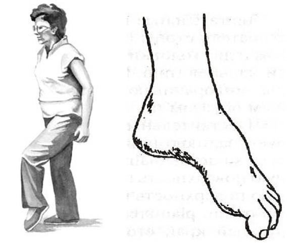
Most often, such disorders appear in the legs, with protrusions and hernias of the lumbar spine.There are motor structures that innervate the muscles of the legs and lower legs.Therefore, with advanced and complicated lumbar osteochondrosis, the legs can beat.It turns inward, the person is forced to lift his leg high to step with the beaten leg, this is called steppage, "cock gait."
But the whole danger of movement disorders is that they can be isolated and not accompanied by pain.And if a person "doesn't have pain," then he may not get to the doctor in time.Therefore, it is very important for patients with progressive protrusions and hernias, for example, the lumbar region, to regularly walk on their toes and heels, and monitor the work of their muscles.
Local symptoms: main signs
Now let's consider the symptoms and characteristics of specific syndromes of cervical, thoracic, and lumbar osteochondrosis.Let's go from top to bottom, from the cervical region to the bottom, through the thoracic region, to the lumbosacral region.
Diagnosis of osteochondrosis
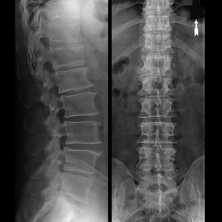
In normal cases, osteochondrosis of the cervical spine and cervical-thoracic occurs as described above.Therefore, the main stage of diagnosis is and remains the identification of the patient's complaints, establishing the presence of concomitant muscle spasms using simple palpation of the muscles along the spinal column.Is it possible to confirm the diagnosis of osteochondrosis using an x-ray examination?
"X-ray" of the cervical spine, and even with functional tests for flexion and extension, does not show the cartilage, because their tissue transmits X-rays.Despite this, based on the location of the vertebrae, one can make a general conclusion about the height of the intervertebral disc, the general straightening of the physiological curvature of the neck - lordosis, as well as the presence of marginal growth on the vertebra with prolonged irritation on its surface by the fragile and dehydrated intervertebral disc.Functional testing can confirm the diagnosis of instability in the cervical spine.
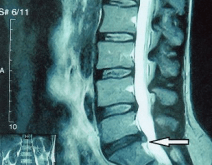
Since the disc itself can only be seen using CT or MRI, magnetic resonance and x-ray computed tomography are indicated to clarify the internal structure of cartilage and formations such as protrusions and hernias.Therefore, with the help of this method, the diagnosis is made accurately, and the tomography results are an indication, as well as a topical guide, for the surgical treatment of hernia in the neurosurgery department.
Treatment of complications of osteochondrosis
Let's repeat once again that it is impossible to cure osteochondrosis, like planned aging and disk dehydration.You can't let things get complicated:
- if there are symptoms of narrowing of the height of the intervertebral disc, then you need to move correctly, do not gain weight and avoid the appearance of protrusions and muscle pain;
- if you already have a protrusion, then you need to be careful not to let it break the fibrous ring, that is, not to turn the protrusion into a hernia, and to avoid the appearance of the protrusion at several stages;
- if you have a hernia, then you need to monitor it dynamically, do a regular MRI, avoid increasing its size, or carry out modern minimally invasive surgical treatment, because without exception, all conservative methods of treating exacerbations of osteochondrosis leave the hernia in place, and only eliminate temporary symptoms: inflammation, pain, shooting and muscle spasms.
But with the slightest violation of the regime, with heavy lifting, hypothermia, injuries, weight gain (in the case of the lower back), the symptoms return again and again.We will explain how you can overcome unpleasant sensations, pain, and limited mobility in the back against the background of exacerbation of osteochondrosis, and existing protrusion or hernia, secondary to social tonic syndrome.
What to do during an exacerbation?
Since there is an attack of acute pain (for example, in the lower back), then you need to follow the following instructions at the pre-medical stage:
- completely eliminate physical activity;
- sleeping on a hard surface (orthopedic mattress or a hard sofa), eliminating the sagging in the back;
- it is advisable to wear a semi-rigid corset to avoid sudden movements and "distortion";
- You should place a massage pillow with a plastic needle applicator on your lower back, or use a Lyapko applicator.You need to keep it for 30 - 40 minutes, 2 -3 times a day;
- after this, ointments containing NSAIDs, ointments with bee or snake venom can be applied to the lower back;
- after rubbing, on the second day you can wrap your lower back in dry heat, for example, a belt made of dog fur.
A common mistake is to warm up on the first day.This can be a heating pad, bathing procedure.At the same time, the swelling only increased, and the pain along with it.You can warm up only after the "highest pain point" has passed.After this, the heat will increase the "absorption" of the swelling.This usually happens in 2-3 days.
The basis of any treatment is etiotropic therapy (elimination of the cause), and pathogenetic treatment (affecting the mechanism of the disease).It is accompanied by symptomatic therapy.For vertebrogenic pain (caused by problems in the spine), things go like this:
- To reduce muscle and spinal swelling, a salt-free diet and limiting the amount of fluid consumed are indicated.You can also give mild potassium-sparing diuretic tablets;
- in the acute phase of lumbar osteochondrosis, short-term treatment can be carried out with intramuscular "injections" of NSAIDs and muscle relaxants: daily.This will help relieve swelling of nerve tissue, eliminate inflammation, and normalize muscle tone;
- in the subacute period, after overcoming the maximum pain, "injections" no longer need to be taken, and attention should be paid to restorative agents, for example, modern drugs of group "B".They effectively restore impaired sensitivity, reduce numbness and paresthesia.
Physiotherapeutic measures continue, the time has come for exercise therapy for osteochondrosis.Its task is to normalize blood circulation and muscle tone, when swelling and inflammation have subsided, but muscle spasms have not completely resolved.
Kinesiotherapy (movement treatment) involves doing therapeutic exercises and swimming.Gymnastics for osteochondrosis of the cervical spine is not aimed at the disc at all, but at the surrounding muscles.Its task is to relieve tonic spasms, increase blood flow, and also normalize venous outflow.This leads to a decrease in muscle tone, a decrease in the severity of pain and stiffness in the back.
Exercises for osteochondrosis must be carried out after a light general warm-up, on "warming up muscles".The main therapeutic factor is movement, not the degree of muscle contraction.Therefore, to avoid repetition, the use of weights is not allowed;gymnastic mats and gymnastic sticks are used.With their help, you can effectively restore range of motion.
Rubbing in the ointment and using the Kuznetsov applicator continues.Swimming, underwater massage, Charcot shower are indicated.It is during the fading aggravation stage that home magnet therapy and physiotherapy are indicated.
Usually treatment takes no more than a week, but in some cases, osteochondrosis can manifest itself with such dangerous symptoms that surgery may be required, and immediately.
About Shants collar
In the initial stage, during the acute stage, it is necessary to protect the neck from unnecessary movement.Shants collars are great for this.Many people make two mistakes when buying this collar.They do not choose it according to their size, which is why it does not perform its function and causes discomfort.
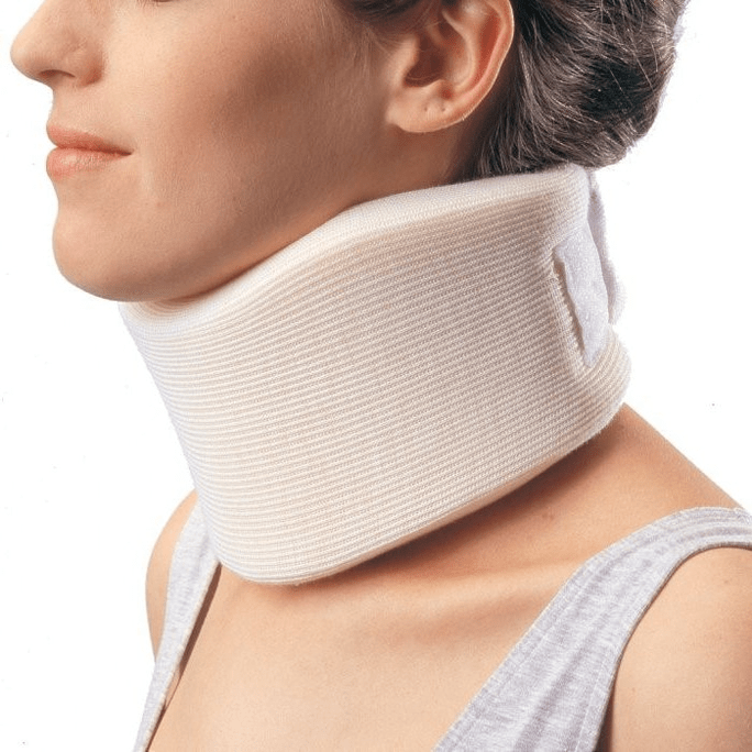
The second common mistake is to wear it for prophylactic purposes for a long time.This leads to weak neck muscles, and only causes more problems.For the collar, there are only two signs where it can be worn:
- the appearance of acute pain in the neck, stiffness and pain spreading to the head;
- if you are going to do physical work in a completely healthy state, where there is a risk of "straining" your neck and getting aggravation.This is, for example, repairing the car, when you lie under it, or washing the window, when you have to reach out and take an awkward position.
The collar should be worn no more than 2-3 days, because wearing it longer can cause venous congestion in the neck muscles, in time to activate the patient.An analogue of the Shants collar for the lower back is a semi-rigid corset purchased at an orthopedic salon.
Surgical treatment or conservative measures?
It is advised that every patient, after the development of symptoms, in the presence of complications, undergo an MRI and consult a neurosurgeon.Modern minimally invasive surgery makes it possible to safely remove relatively large hernias, without prolonged hospitalization, without being forced to lie down for several days, without affecting the quality of life, because it is performed using modern video endoscopy, radio frequency, laser technology or using cold plasma.You can steam part of the kernel and lower the pressure, reducing the risk of getting a hernia.And you can eliminate the defect radically, that is, by getting rid of it completely.
There is no need to be afraid to operate on a hernia;this is no longer the previous type of open operation in the 80s-90s of the last century with muscle surgery, blood loss and a long recovery period.They are more like small punctures under X-ray control followed by the use of modern technology.
Prevention of osteochondrosis and its complications
Osteochondrosis, including the complicated ones, the symptoms and treatment of which we discussed above, is largely not a disease at all, but only an inevitable manifestation of aging and premature "shrinkage" of the intervertebral disc.Osteochondrosis needs little to not bother us:
- avoid hypothermia, especially in autumn and spring, and fall in winter;
- do not lift loads, and carry loads only with a straight back, in a backpack;
- drink more clean water;
- don't be fat, your weight should match your height;
- treating flat feet, if any;
- do physical exercise regularly;
- engage in a type of exercise that reduces the burden on the back (swimming);
- abandon bad habits;
- replace mental stress with physical activity.After every hour and a half of mental work, it is recommended to change the type of activity to physical work;
- You can regularly do at least an x-ray of the lumbar spine in two projections, or an MRI, to find out if a hernia, if any, is developing;
By following these simple suggestions, you can keep your back healthy and mobile for life.